Friday 25 February 2022

Lupine publishers | Crystallization and Polymorphism-Scalable Process for Celecoxib and It’s Polymorph From-3 (Non- Steroidal Anti-Inflammatory Drug (NSAID)

 Lupine Publishers | Crystallization and Polymorphism-Scalable Process for Celecoxib and It’s Polymorph From-3 (Non- Steroidal Anti-Inflammatory Drug (NSAID)


Abstract

The present process provides an improved process for the preparation of 4-[5-(4-methylphenyl)-3- (trifluoromethyl)-1Hpyrazol- 1-yl] benzene sulfonamide (Celecoxib) and its purification and crystallization to produce polymorph. The present process, which describes the manufacturing process of Celecoxib, which is a non- steroidal anti-inflammatory drug (NSAID), has the advantage of scaling up to the industrial level of production. The process uses safe reagents in the process which makes it for industrial scale operations. The yields in the process are high, which makes it a cost-effective process. Formation of isomers are less compared with the all existing process, which makes it effective to make it to the pharmacopoeia grade. Residual solvents play a very important role in the impurity profile of APIs as per the ICH Guidelines ICH Q3C (R4). In this process by carrying out the final step of condensation in the aqueous medium followed by crystallization, the residual solvents limits are well taken care of.

Keywords: Non-steroidal anti-inflammatory drug (NSAID); Celecoxib; Cyclooxygenase 2; X-ray diffraction; Polymorphism; Process

Discussion

Figure 1: Classes of multi component molecular crystals.


Figure 1 present process relates to “AN IMPROVED PROCESS FOR THE PREPARATION OF CELECOXIB POLYMORPH FORM”. Celecoxib is designated chemically as 4-[5-(4-methylphenyl)-3- (trifluoromethyl)- 1H-pyrazol-1-yl] benzene sulphonamide and is a diaryl-substituted pyrazole [1]. The compound has the following structure (Figure 2).

Figure 2: 4-[5-(4-methylphenyl)-3-(trifluoromethyl)-1Hpyrazol- 1-yl] benzene sulfonamide.


The drug is currently marketed as Celebrex® in the United States of America by Pharmacia Corporation. Celecoxib is a non-steroidal anti-inflammatory drug (NSAID) [1] mainly used in treatment of arthritis, pain, menstrual cramps, and colonic polyps. Celecoxib blocks the enzyme (cyclooxygenase 2) which makes prostaglandins, resulting in lowering the concentrations of prostaglandins. As a consequence, reduction in inflammation and its accompanying pain, fever, swelling and tenderness. The manufacture of Celecoxib has been described in various patents and to cite a few references, G. D. Searl & Co. has disclosed method for preparation of Celecoxib [2-3] in US 5,466,823 which is as under: US 5,134,142 [2], US 5,563,165, US 6,150,534, US 5,892,053, US 2007/0004924, US 2008/0234491, EP 1,528,058, EP 1,167,355, EP 2,246,332, WO 01/42221, WO 03/090730, WO05/014546, WO 06/051340, WO 08/145733, and WO 2010/095024 have also described the synthesis of Celecoxib Reddy et al in their publication in Org. Process Res. Dev., 2009, 13(1), pp 98-101. have disclosed the synthesis (Figure 3).

Figure 3: Manufacture of Celecoxib.


Detailed Description of the Drawings

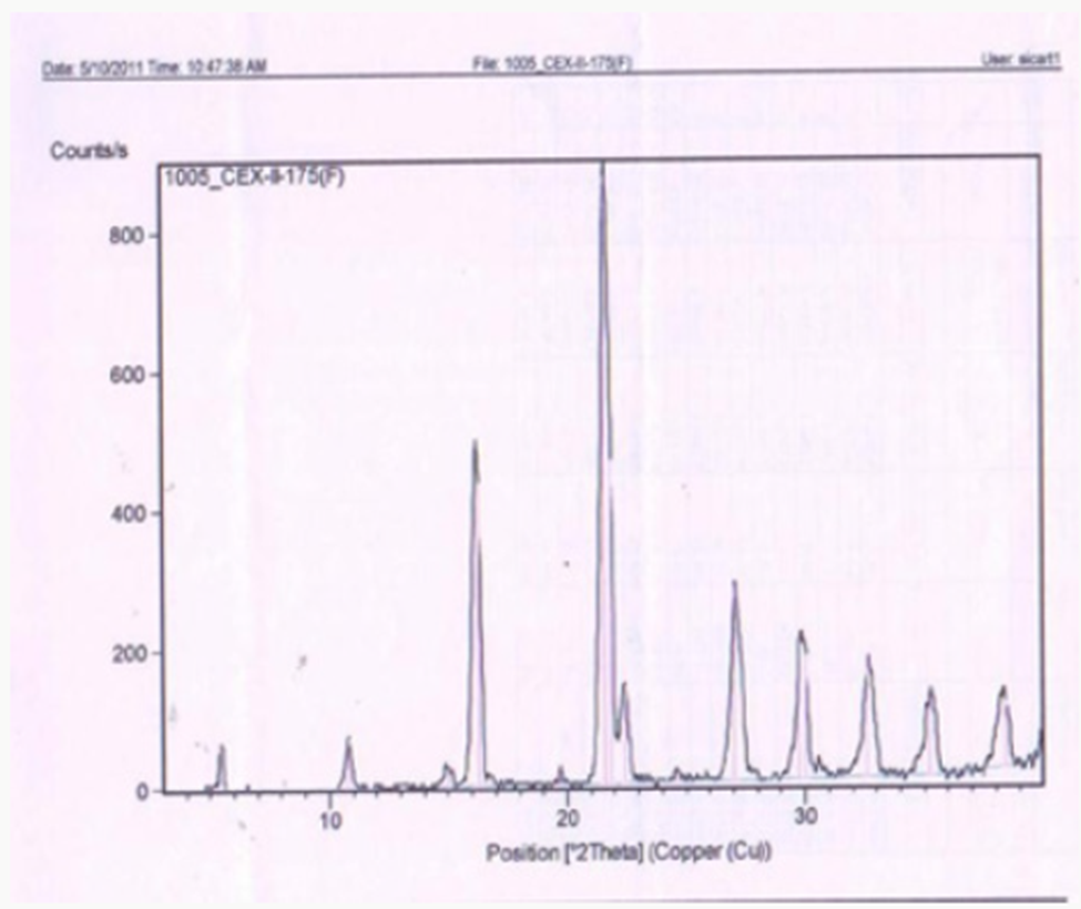
Figure 1 describes the powder X-ray diffraction pattern of the Celecoxib Polymorph; Figure 2 illustrates 2θvalues. Figure 3 depicts the DSC thermogram taken at 10:C /min over a temperature range of 30:C to 200:C for Celecoxib polymorphic form.

Description of the Process

Figure 4: Crystals of Celecoxib polymorph.


The present procedure describes the preparation of Celecoxib by a novel process and its crystallization to polymorphic form. The present process for the preparation of Celecoxib by a process involving condensation of 4,4,4-trifluoro-1-[4-(methyl) phenyl]- butane-1,3-dione [1] with sulphonamido phenyl hydrazine hydrochloride [2] in an aqueous medium to give Celecoxib [3]. This is followed by crystallization from a mixture of solvents [4- 8] containing Aromatic hydrocarbon and aliphatic ketone. In the condensation reaction the reactants are added in water and reactions done at ambient temperature. The crude Celecoxib is isolated by filtration. In the for purification of Celecoxib and its crystallization to polymorphic FORM Preparing a solution of Crude Celecoxib in a solvent mixture comprising of an aliphatic ketone (Acetone) and an aromatic hydrocarbon (Toluene)at reflux temperature followed by cooling crystallization to give crystals of Celecoxib polymorph [8-12] (Figure 4).

Table 1:


In this process by carrying out the final step of condensation in the aqueous medium followed by crystallization, the residual solvents limits are well taken care of. The yields in the process are higher compared to the prior art, which makes it a cost-effective process. Formation of isomers are less compared with the prior art, which makes it effective to make it to the pharmacopoeia grade. Residual solvents play a very important role in the impurity profile of APIs as per the ICH Guidelines ICH Q3C (R4). In this process by carrying out the final step of condensation in the aqueous medium followed by crystallization, the residual solvents limits are well taken care of [13,14]. The crystallization conditions are well established to give crystalline polymorph. The powder X-Ray diffraction pattern of the Celecoxib is given in Figure 1 and 2θ values are given in Table 1 of Figure 2. The differential scanning calorimeter graph of the Celecoxib polymorph under specific conditions shows the melting point around 162.7˚C. The DSC of Celecoxib is given in Figures 3,5, and 6.

Figure 5: Solid Dosage Forms.


Figure 6: Process induced transformations.


Solid Forms

a) Propensity to produce different forms not significantly different for salts and non-salts.

b) Need more data on co-crystals (Figure 7).

The details of the new methods for preparation of celecoxib are further illustrated in the following examples.

Example 1: Preparation of Celecoxib

In a 20 liter 3-necked flask, equipped with stirrer, thermometer and reflux condenser, deionized water (7.9 Liter) is charged and mixture of 4,4,4-trifluoro-1-[4-(methyl) phenyl]-butane-1,3-dione (1.6Kg; 6.95×10³mmoles) and 4-sulphonamido phenyl hydrazine hydrochloride (1.7Kg; 7.57×10³mmoles), a resultant mixture was heated at 75˚C to 80˚C and maintained for 5 hours. The reaction mixture was cooled to 25˚C to 30˚C to give a slurry. The slurry was filtered and washed with water (3.2liter) wet- cake was collected and further processed for purification as given below.

Figure 7: Percentages of forms from Polymorph Screening.


a) Purification and Crystallization to Give Polymorph: Celecoxib wet-cake obtained in the process described above was taken into 20 liter 3-necked flask, equipped with stirrer, thermometer and reflux condenser, mixture of acetone (0.54liter) and toluene (10.8liter) was added and the reaction mixture was heated to 80˚C to 85˚C for 30 minutes. Activated carbon (0.3Kg) was added and the reaction mixture was further heated to 80˚C to 85˚C. The reaction mixture was cooled to 25˚C -30˚C. The slurry was filtered, washed with toluene and then dried at 70˚C to yield the Celecoxib polymorph compound1.35 kg (HPLC purity-99.8% & molar yield; 50.9%).

IR: 3340, 3240, 1600, 1500, 1350, 1280, 1235, 1160, 980, 910, 840, 800,760, 635, 560, 530 cm-1 (KBr pellet)

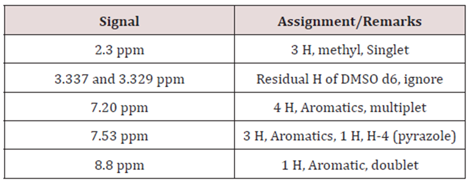
Proton NMR: Solvent: DMSO d6, 300 MHz.

Example 2: Preparation of Celecoxib

In a 20liter 3-necked flask, equipped with stirrer, thermometer and reflux condenser, charge deionized water(9Liter) and mixture of 4,4,4-trifluoro-1-[4-(methyl)phenyl]-butane-1,3-dione(1.6Kg; 6.95×10³mmoles) and 4-sulphonamido phenyl hydrazine hydrochloride(1.7Kg; 7.57×10³mmoles), a resultant mixture was heated at 90˚C to 100˚C and maintained for 5 hours. The reaction mixture was cooled to 25˚C to 30˚C. The slurry was filtered and washed with water (3.2liter) wet-cake was collected and further processed for purification as given below.

Figure 8: Powder X-Ray diffraction pattern of the Celecoxib.


a) Purification and crystallization to give Polymorph: Celecoxib wet-cake obtained in the process described above was taken into 20liter 3-necked flask, equipped with stirrer, thermometer and reflux condenser, mixture of acetone (0.54liter) and toluene (10.8liter) was added and the reaction mixture was heated to 80˚C to 85˚C for 30 minutes. Activated carbon (0.3Kg) was added and the reaction mixture was further heated to 80˚C to 85˚C. The reaction mixture was cooled to 25˚C -30˚C. The separated solid was filtered, washed with toluene and then dried at 70˚C to yield the Celecoxib polymorph compound1.24 kg (HPLC purity-99.3% & molar yield; 47%) (Figures 8-11c).

Figure 9: 2θ values.


Figure 10: DSC of Celecoxib.


Figure 11c:


Conclusion

The distinct advantage of the present method of preparation over the prior art can be summarized as per below:

The present process, which describes the manufacturing process of Celecoxib, which is a non- steroidal anti-inflammatory drug (NSAID), has the advantage of scaling up to the industrial level of production. The process uses safe reagents in the process which makes it for industrial scale operations. The present process provides an improved process for the preparation of 4-[5-(4-methylphenyl)-3- (trifluoromethyl)-1H-pyrazol-1- yl] benzene sulfonamide (Celecoxib) and its purification and crystallization to produce polymorph. The yields in the process are high compared to existing process which makes it a cost-effective process. Formation of isomers are less compared with the prior art, which makes it effective to make it to the pharmacopoeia grade. In this process by carrying out the final step of condensation in the aqueous medium followed by crystallization, the residual solvents limits are well taken care of. The yields in the process are higher compared to the prior art, which makes it a cost-effective process. Residual solvents play a very important role in the impurity profile of APIs as per the ICH Guidelines ICH Q3C (R4).

Read More Lupine Publishers Medical Sciences Articles: https://lupine-publishers-medical-sciences.blogspot.com/

Sunday 20 February 2022

Lupine Publishers | Defining Satellite Robotics Surgery using IOT

 Lupine Publishers | Defining Satellite Robotics Surgery using IOT


Opinion

Now days Internet of Things (IoT) is making everything, remote control and remote operating possible and change imagination of objects communication into reality using Satellite based USN (Ubiquitous Sensing Network). IoT is all ultimate communication technology where not only living but also all non-living things can communicate, command, control, process using their unique RFIDs and USN. Hence it would be possible what I hypothesis “Satellite Robotic Surgery using IoT”. I have drawing one model to explain how this happen will possible in near future labeled as “Satellite Robotics Surgery Model (SRSM)”. Let me explain you how it would be engineer and functional (Figure 1).

To implement Satellite based robotics surgery using IoT very first requirement is Various Medical Surgeries Subroutine/ Programs/Templates which passes through Medical Intelligence System to decide which surgery procedure requested from client hospital from which country and what surgical method is efficient from the alternatives subroutines and what are seriousness, complication and nature of surgery. After medical intelligence decision support system decision commands prepared and send to command processing unit. The function of command processing to caliber command with precise control, time management, signal conditioning and data acquisitions. At next level whole process included its RFID and streaming through transmission unit to client’s hospitals from 1, 2, 3 … Nth using USN and IoT with satellite-based communication worldwide with granting to requests of number of client’s hospital who requested for satellite based robotic surgery using IoT.

Conclusion

I have discussed how Satellite Robotic Surgery possible using IoT and USN with the help of Satellite Robotics Surgery Model (SRSM)”. The big advantage of this technology surgical operation possible from expert programs with absence of doctors but one big disadvantage would be if data streaming command communication failure or break at any point become cause of stop remote surgery or obstacle because of distortion in signal reception at client’s hospitals.

Acknowledgment

I really thankful to my wife Safeena Shaikh for her moral support my sons Md. Nameer Shaikh & Md. Shadaan Shaikh for their love which keeps me fresh with new ideas and my close friend Tanvir Sayyed for her positive support with me. I acknowledge this work to my friends Jyoti Firke and Ritashri Cahudhari for encouragement and equally to Dr. BN Gupta who inspired me.

Read More Lupine Publishers Medical Sciences Articles: https://lupine-publishers-medical-sciences.blogspot.com/

Saturday 5 February 2022

Lupine Publishers | Final Solution in Femoroacetabular Impingement - Threaded Cups in Hip Revision Surgery 2002-2012

 Lupine Publishers | LOJ Medical Sciences


Abstract

Purpose: Here, we report the complications of endoprosthesis with threaded cups according to our application in cases of complicated acetabulum (hip revisions and femoroacetabular impingement (FAI)).

Methods: A total of 504 patients was analyzed, including 189 men and 315 women. For re-implanted patients (n=49), the mean time to re-implantation was 6.3 years and the average age at re-implantation was 54.7 years. For censored patients (n=455), the mean time to censoring was 5.5 years and the average age of the implant was 55.7 years. Among the patients that were censored, 77 died and 378 patients did not experience an event necessitating re-implantation.

Results: For the total study population, the Kaplan-Meier estimate of 5-year survival is 0.94 and of 10-year survival is 0.85. 90% of the implants survived 7 years, 80% of the implants survived almost 12 years and 70% of implants survived almost 15 years. Kaplan-Meier survival analysis suggests that men have higher survival than women. Age of the patient at the time of implant was not a statistically significant factor for re-implantation (p value=0.21) but sex was (p value=0.02). Women had 2.25 times more risk of being re-implanted than men.

Conclusion: In our case series, the failure rate, as a result of aseptic loosening, was 7.4% (n=51). More than half the failure cases (56%) required re-implantation. Over one-third (37%) of the primary arthrosis cases, were found to be FAI, lessening the frequency of diagnosis of the former. In our clinic, the threaded cup seems to be indispensable in hip revision surgery and in treating FAI.

Keywords: Hip; FAI; Conclusion

Acknowledgements
This article does not contain any studies with human participants or animals performed by any of the authors.

Abstract


In our hospital, the most common indication for hip revision surgery is post dysplastic arthrosis, followed by femoroacetabular impingement (FAI), early progressive and deformative arthrosis of the hip joint and protrusio acetabuli, with cases of achondroplasia or deformative arthrosis in hereditary multiple exostoses being rarer but not uncommon. The cases of post dysplastic arthrosis represent developmental dysplasia of the hip (DDH), epiphyseal dysplasia, spondylo-epiphyseal dysplasia, and proximal femoral focal deficiency (PFFD) or neurogenic luxation of the hip. In our experience, a small portion of the patients requiring hip revision surgery have shown necrosis of the femoral head, manifesting from chondrolysis of the hip joint, from coxitis (including tuberculosis coxitis), from radiation therapy and from hemophilia. In addition, we noted over time, that a small portion of rheumatic patients were treated with the use of threaded cups.

In our experience, we have also noted that a portion of the patients who were treated with threaded cups experienced complications. The complication of posttraumatic arthrosis has not been uncommon, the conditions of which involve slipped capital femoral epiphyisis, acetabular fractures either treated conservatively or operatively, pseudoarthrosis after femoral neck fractures or fusion of the hip joint. Cancer patients have represented a distinctive group for postsurgical complications (including need for re-implantations). The types of cancers related to the original hip revision surgeries have been benign acetabular cysts, Hodgkin’s lymphoma, Ewing’s sarcoma, and giant cell tumor or desmoplastic fibroma in the supra-acetabular region. In our hospital, the ratio of primo-implantation to total implantations has been relatively low and that of re-implantations has been nearly one-third, making threaded cups irreplaceable in hip revision surgery at our institution.

In order to share the knowledge from our collective experience with hip revision surgery using threaded cups for diverse cases, we performed a systematic evaluation of the overall complications our patients have experienced from hip revision over a 10-year period (2002-2012).

Materials and Methods


In the 10-year period of 2002-2012, a total of 565 patients underwent hip revision surgery with threaded cups. Of those, 504 patients had complete data and were selected for analysis (189 men and 315 women). The total study population included 49 patients with re-implantation and 455 patients that were censored. For the re-implanted patients, the mean time to re-implantation was 6.3 years and the mean age at the time of implant was 54.7 years. For the censored patients, the mean time to censoring was 5.5 years and the mean age at implant was 55.7 years (Table 1). Among the censored patients, 77 died and 378 did not experience an event necessitating re-implantation.

Table 1: Descriptive statistics of time to event and age of the patient at the time of implant for the study population. Data are presented in years.


Among the total study population, 16.8% had indication for primary arthrosis. In general, those patients were relatively younger and with early progressive and deformative arthrosis of the hip joint, hip arthrosis with protrusion, and FAI. We also recorded the numbers of achondroplasia patients or deformative arthrosis in hereditary multiple exostoses. When we selected 100 patients from among the group of primary arthroses cases, 37% were found to be FAI, indicating that FAI is secondary arthrosis in origin.

Figure 1: Representative case of high iliac luxation – Hartofilakidis Type III, Crowe Type IV, Eftekhar Type D hip dysplasia, solved with bilateral endoprosthesis (THR) and having visible difficulties with stem implantations.


Patients with post dysplastic arthrosis were the largest group among the hip revision surgery cases (60.3%). These cases included DDH, epiphyseal dysplasia, spondylo-epiphyseal dysplasia, and PFFD or neurogenic luxation of the hip representing nearly every type of difficult anatomical acetabulum for restoration of true acetabular region or for re-lengthening of the lower extremities (Figure 1). Among the total study population, 10.4% had indication of necrosis of the femoral head. These cases consisted of such conditions as chondrolysis of the hip joint, following coxitis (including the stages after tuberculosis coxitis), or necrosis resulting from radiation therapy or in relation to hemophilia (Figure 2).

Figure 2: A patient requiring hip revision surgery following septic coxitis with necrosis of the head of the femur. The patient underwent THR. (a) 3-months post-operative. (b) 6-months post-operative. (c) Aseptic loosening of the cup detected at 9 months follow-up. (d) Re-implantation with larger cup.


Rheumatic patients in our study population had received longterm corticoid therapy; among these patients, osteoporosis and necrosis of the femoral head is common. The threaded cup was used in 3.2% of this study subgroup. The rheumatic patient group also included several cases of ankylosing spondylitis (also known as Bechterev’s disease) and juvenile rheumatoid arthritis. Regarding the difficult anatomical acetabulum of these cases, 7.7% of the patients implanted with threaded cups experienced posttraumatic arthrosis, the conditions of which involved slipped capital femoral epiphyisis, acetabular fractures either treated conservatively or after osteosynthesis, pseudoarthrosis after femoral neck fractures or conditions after posttraumatic arthrodesis of the hip joint.

Among the total study population, rare indication was oncological status (1.6%). Such cases consisted of benign acetabular cysts, Hodgkin’s lymphoma, Ewing’s sarcoma, giant cell tumor or desmoplastic fibroma in the supraacetabular region. When we compare threaded THR primo-implantations to total number of primo-implantations at our institution in the 10-year study period, there was an average 13.1% of primo-implantations. The same comparison for the re-implantation group showed that we used the threaded THR 27.2% on average, making this treatment modality indispensable in hip revision surgery at our institution.

Figure 3: Representative case of cup breakage due to loss of bone in the supero-lateral part of the acetabulum affected with metastatic process.


We noticed aseptic loosening of the cup in 48 cases, of which 18 cups loosened after primo-implantation and 29 loosened after re-implantation, giving a failure rate of over one-half (56%). This high percentage is largely due to the area of implantation being technically difficult, which necessitated the use of a different device (i.e. the threaded cup); the rest represented failure of the cups after acetabular fractures (2 cases) and after metastatic affections surrounding the acetabulum (2 cases). Dislocations of the THR occurred in 4.4% (30 cases) of the study population, and 15 of the cups required operative intervention. In 5 cases, open reduction was enough; in 9 cases we re-implanted the inlay or head with one of a bigger size, and in 6 cases we performed re-implantation of the stem or cup to correct the position of the implant.

There were infectious complications in 3% (21 cases) of the study population. In 17 of those cases, a revision procedure was required, including 5 revisions to address an early superficial infection and 2 revisions for deep infection. Proper drainage with retained implant was achieved in all cases. A total of 13 patients remain in follow-up to date, including 3 patients with chronical fistulation, 8 patients who underwent two-stage re-implantation of the THR, and 2 patients who underwent implant removal and had no further re-implantation.

There was slow migration of the implant into the pelvis or up into the iliac bone without obvious loosening in 2.3%. This migration is likely explained by loosening of the circumferential grip of the cortical bone, when the anterior and/or posterior acetabular wall is weakened. In such a condition of cortical weakening, the underlying cancellous bone does not have enough mechanical strength and thus the cup migrates (Figure 4).

Figure 4: Representative case of slow migration up into iliac bone, with loosening at 8 years after the primary implantation.


Graph 1: Kaplan-Meier survivor analyses.


Considering the threaded cup concept, we have observed stress shield effect often. Since the stress shield exerts its effect in cup migration, focused follow-up is necessary to obtain meaningful findings. The advantage of a threaded cup is that there is no need of full coverage. In particular, 70% of coverage is sufficient, without need of augmentation [1]. In addition, arthritis with protrusion (FAI pincer type) is one of the indications for the threaded cup since those cases do not need support of the acetabular floor. Implantrelated complications are to be considered as neuro-vascular complications. Among our study population, there was a total number of 10 nerve palsies (1.5%), 4 of which had to be operated; two of those patients underwent sural graft transplantation due to femoral nerve cutoff, [2] and the other two underwent the same procedure for static nerve palsy, with one requiring simultaneous anterior tibial tendon transfer. In one case, we released the lateral cutaneous nerve (Graph 1). The Kaplan-Meier survical curve suggests that men have higher survival than women do (Graph 2).

Graph 2: Kaplan-Meier survivor curve for men and women, showing the association of time to event to sex.


Age of the patient at the time of implantation was not statistically significant (p value = 0.21). Sex was a statistically significant factor (p value 0.02). Women had 2.25 times more risk of being re-implanted than men (Table 2).

Table 2: Cox model of proportional hazard for patient sex, showing dependence of time to event with age at the implant and sex.


Conclusion

 Aseptic loosening reportedly occurs in around 6.5% of implanted patients at 4 years following placement [3]. In one study the radiological sign of instability was found in 21% of patients (n =130) at a mean follow-up of 3.9 years [4]. In another study [5], 17 cups (accounting for 25% of the cases assessed) were revised at an average of 5.2 years, with another 9 additional cases awaiting revision. Brujin et al. [2] reported migration for 25% of threaded cups in a follow-up of 4.5 years, and a 6% revision rate. It has been suggested that high and continuous stress near the threads may not allow bony in-growth [6] and may produce ischemia and necrosis [7] that require surgical revision in 14 months following the implantation surgery [8].

Discussion

Overall, 7.4% of our patients experienced loosening of the threaded cups and 3% experienced infectious loosening, which agrees with the literature [9,10]. In general, loosening of threaded cups does not give a good clinical and/or radiological outcome over the long term. X-ray appearance deteriorates with time, without evidence of an initial clinical correlation. The appearance of relative lines does not necessarily equate to the occurrence of loosening, but such a finding should be monitored carefully thereafter. Detection of osteolysis, however, indicates that loosening will occur [11]. A study by Harris et al from 1986 indicated that 79% of coxartosis cases originated from the cam impingement, with the rest by rheumatoid arthritis (21%) and hemochromatosis (10%).

In our series, when we compared 100 patients admitted for the THR procedure, 37% showed FAI signs, with the average age of implantation being 58 years old. There were 23% post-DDH cases, 11% idiopathic avascular necrosis, 17% post-trauma cases, and 12% others; thus, the diagnosis of primary arthrosis is much less common. FAI both types are finally resolved by a THR and indicated much earlier than primary arthrosis. As changing in orientation of the acetabuli we use these threaded cups, [12] more in pincer type of the FAI, where orientation is essential. We don’t necessarily use these cups, though in special cases we have to prefer primary stability to have good long-term results. The failure of the threaded acetabular component is due to difficulties of anatomy of the acetabulum, as observed in the cases in which we used this component.

Even though we decided to use this special design of threaded cup, despite its higher rate of complications, we were able to resolve one-third of our re-implantation cases and to overcome the difficulties of the acetabulum in FAI [13,14]. Kaplan-Meier survival curve analyses identified threaded cups as a good option (with survival of 10 years); the Cox proportional curve indicated higher risk for re-implant for women. Even though we investigated THR as a last option for patients with FAI, pelvic transfers or any cervicocapital remodeling may also represent last options. Ultimately, this study did not show the threaded cup to be ideal for complicated acetabulum, though in our clinic it is ideal for this diagnosis.

Read More Lupine Publishers Medical Sciences Journal Articles: https://lupine-publishers-medical-sciences.blogspot.com/


The Body Electric: Humans Have A ‘Force Field’ Around Their Bodies

  Abstract Bioelectronic medicine (BEM) is the most recent medical revolution — not an innovation or an improvement or a step up but a radic...