Showing posts with label international journal of medical sciences. Show all posts

Monday, 22 November 2021

Lupine Publishers| Diagnosis and Immunotherapy Strategies for Melanoma: A Review

 Lupine Publishers| Journal of Medical Sciences


Abstract

Melanoma is a type of skin cancer. Melanoma skin cancer is made up of abnormal pigment cells (melanocytes). Melanocytes are located at the bottom of the epidermis. These cells make melanin, which spreads to the top of the epidermis and gives skin its colour. Melanoma is usually brown or black in colour because the cell still makes melanin. Melanoma is more dangerous than other skin cancers because it more likely to spread if it is not found early. However, most melanomas about 84 out of 100 are found early before they have spread and so are likely to be cured with treatments. Melanoma can occur on any skin surface. In men, it’s often found on the skin on the head, on the neck, or between the shoulders and the hips. In women, it’s often found on the skin on the lower legs or between the shoulders and the hips. Melanoma is rare in people with dark skin. When it does develop in people with dark skin, it’s usually found under the fingernails, under the toenails, on the palms of the hands, or on the soles of the feet. Although one of the less common types of skin cancer, melanoma is considered the most serious type of skin cancer because it is more likely to spread to other parts of the body, especially if not detected early. The earlier melanoma is found, the more successful treatment is likely to be. Despite the many investigations in this field and a rapidly growing knowledge base, classification according to specific mutational profiles is not yet validated. Further investigations are required for validation and refinement, and to possibly identify additional factors.

Keywords: Melanoma; Melanocytes; Epidermis; Melanin

Introduction

Cancer is a disease of the cells, which are the body’s basic building blocks. The body constantly makes new cells to help us grow, replace worn-out tissue and heal injuries. Normally, cells multiply and die in an orderly way. Sometimes cells don’t grow, divide and die in the usual way. This may cause blood or lymph fluid in the body to become abnormal, or form a lump called a tumour. A tumour can be benign or malignant.

Benign tumour

Cells are confined to one area and are not able to spread to other parts of the body. This is not cancer.

Malignant tumour

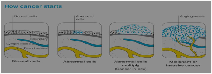
Figure 1: How cancer starts?
Lupinepublishers-openaccess-biomedicalengineering-biosciences

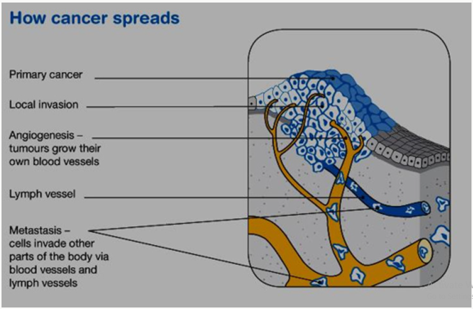
This is made up of cancerous cells, which sometimes also have the ability to spread by travelling through the bloodstream or lymphatic system (lymph fluid) (Figure 1). The cancer that first develops in a tissue or organ is called the primary cancer. A malignant tumour is usually named after the organ or type of cell affected. A malignant tumour that has not spread to other parts of the body is called localized cancer. A tumour may invade deeper into surrounding tissue and can grow its own blood vessels in a process called angiogenesis. If cancerous cells grow and form another tumour at a new site, it is called a secondary cancer or metastasis. A metastasis keeps the name of the original cancer. For example, melanoma that has spread to the bones is called metastatic melanoma, even though the person may be experiencing symptoms caused by problems in the bones (Figure 2).

Figure 2: How cancer spreads?
Lupinepublishers-openaccess-biomedicalengineering-biosciences

The skin

The skin is the largest organ in the body. It acts as a barrier to protect the body from injury, control body temperature and prevent loss of body fluids. Skin, like all other body tissues, is made up of cells. The two main layers of the skin are the epidermis and the dermis. Below these is a layer of fatty tissue (subcutis). The epidermis is the top, outer layer of the skin. It contains three main kinds of cells:

Squamous cells - These flat cells are packed tightly together to make up the top layer of skin and form the thickest layer of the epidermis. These cells eventually die and become the surface of your skin. Over time our body sheds these dead skin cells.

Basal cells: These block-like cells make up the lower layer of the epidermis and multiply constantly. As they age, they move upwards in the epidermis and flatten out to form squamous cells.

Melanocytes: These cells sit between the basal cells of the skin and produce a dark pigment called melanin, the substance that gives skin its colour. When skin is exposed to ultraviolet (UV) radiation melanocytes make extra melanin to try to protect the skin from getting burnt. This is what causes skin to tan. Melanocytes are also in non-cancerous (benign) spots on the skin called moles or naevi. Most moles are brown, tan or pink in colour and round in shape [1].

Melanoma

All cancers involve the malfunction of genes that control cell growth and division. Most cancers are diagnosed in persons 55 and older. The lifetime risk for cancer is approximately 50% for men and approximately 33% for women.1 About 5% to 10% of all cancers are clearly hereditary; the other 90-95% of cancers are not hereditary but result from damage to genes (that is, somatic mutations) due to internal factors (that is, hormones, digestion of nutrients) or external factors (such as tobacco, chemicals, and sunlight). Cancer treatment includes surgery, radiation, chemotherapy, and vaccines. Some cancers are treated using various combinations of surgery, radiation, and chemotherapy [2]. Basal cell carcinoma and squamous cell carcinoma are the most common skin cancers. The most serious common skin cancer is melanoma (malignant melanoma), which arises from neural crest-derived melanocytes located in the epidermis or dermis of the skin. Melanomas also can arise from melanocytes located in other regions of the body such as the eye, meninges, digestive tract, or lymph nodes. Melanocytes in the skin can give rise to a number of benign lesions such as nevi (moles), dysplastic nevi, Spitz nevi, blue nevi, solar lentigo, and seborrheic keratosis. When dealing with melanoma of the skin, establishing which pigmented lesions are malignant is the prime clinical problem, as the vast majority are benign.

Epidemiology and Aetiology

Exposure to the sun is the most commonly associated factor [3,4]. Predisposing factors that have been found to increase the risk of melanoma include skin that sunburns easily, poor tanning response, light-colored skin, history of severe sunburns, numerous nevi (more than 50), atypical nevi (that is, dysplastic nevi), a tendency toward freckling, a history of skin cancer, a family history of melanoma, the use of tanning salon, previous melanoma, and a weakened immune system (due to other cancers, transplant drugs, or HIV infection). Oral methoxsalen (psoralen) and ultraviolet A radiation is an effective treatment for psoriasis, but it is carcinogenic and increases the risk for melanoma. Individuals with recreational and vacation sun exposure may be at greater risk than those whose occupation exposes them to the sun constantly. In fact, continuous sun exposure appears to be a protective factor where melanoma is concerned.

Melanoma is a common cancer. In early 2005, the American Cancer Society estimated that approximately 59,500 individuals would be diagnosed with melanoma, the projected number of deaths was 7,770. The prevalence of melanoma is 13:1,000 Caucasian Americans. During the 1970s, the incidence rate of melanoma showed a marked increase of approximately 6.0% per year, although this rate slowed to approximately 3.0% a year beginning in 1981. There is some data to suggest that superficial spreading melanoma has been found more frequently during the last 30 years. Five-year survival rates for melanoma of the skin have increased from 80% in 1976 to 85% in 1985 to 91% in 2000 [5].

Types of melanoma

Superficial spreading melanoma: It begins with an intraepidermal horizontal or radial growth phase, appearing first as a macule that slowly evolves into a plaque, often with multiple colours and pale areas of regression. Secondary nodular areas may also develop. This is the most common type of melanoma, making up 55-60% of all cases. It is more common in younger people and is often related to a pattern of irregular high sun exposure, including episodes of sunburn. It can start as a new brown or black spot that spreads within the outer layer of the skin (epidermis), or an existing spot, freckle or mole that changes size, colour or shape. It can develop on any part of the body. This type of melanoma often grows slowly and becomes more dangerous when it invades the lower layer of the skin (dermis) [6].

Nodular melanoma

Is a primarily nodular, exophytic brown-black, often eroded or bleeding tumour, which is characterized by an aggressive vertical phase, with a short or absent horizontal growth phase. This type makes up about 10-15% of melanomas. It usually appears as a round, raised lump on the surface of the skin that is often red, pink, brown or black and feels firm to touch. It may develop a crusty surface that bleeds easily. It is most commonly found in older people on severely sun-damaged skin on the head and neck. It is a fast growing and aggressive form of melanoma, spreading quickly into the lower layer of the skin (dermis) [7].

Lentigo maligna melanoma: Arises often after many years from a lentigo maligna (melanoma in situ) located predominantly on the sun-damaged faces of older individuals. This type of melanoma is most common in older people. It makes up about 10-15% of melanomas and begins as a large freckle (lentigo maligna) in an area of sun-damaged skin, such as the face, ears, neck and head. It may grow slowly and superficially over many years before it penetrates more deeply into the skin. Acral lentiginous melanoma: is typically palmoplantar or subungual. In its initial intraepidermal phase (which may be protracted), there is irregular, poorly circumscribed pigmentation; later a nodular region reflects the invasive growth pattern. This is an uncommon type of melanoma (around 1-2% of all cases). It is most commonly found on the hairless skin on the soles of the feet or palms of the hands, or under the fingernails or toenails. It commonly appears as a colourless or lightly pigmented area, which can be mistaken for a stain or bruise. In the nails, it most often presents as a long streak of pigment in the nail or discoloration in the skin around the nail. It may grow slowly before it becomes invasive.

Desmoplastic melanoma

This is another uncommon type of melanoma (around 1-2% of cases) that presents as a firm, progressively growing lump, often on the head or neck. Many are skin-coloured and not pigmented and can be difficult to diagnose. There are some other rarer types of non-skin melanoma. Mucosal melanomas start in the tissues in the mouth, nervous system, anus, urethra, vagina, and nasal passages. Ocular melanomas start in the eye. Recent molecular studies have shown the genetic heterogeneity of melanoma, with distinct molecular signatures identified in tumours at different anatomical locations and with different associations with reported sun exposure.

Symptoms of Melanoma

Often the first sign of melanoma is a change in the shape, color, size, or feel of an existing mole. Melanoma may also appear as a new mole. Thinking of “ABCDE” can help you remember what to look for

a) Asymmetry: The shape of one half does not match the other half.

b) Border that is irregular: The edges are often ragged, notched, or blurred in outline. The pigment may spread into the surrounding skin.

c) Color that is uneven: Shades of black, brown, and tan may be present. Areas of white, gray, red, pink, or blue may also be seen

d) Diameter: There is a change in size, usually an increase. Melanomas can be tiny, but most are larger than the size of a pea (larger than 6 millimeters or about 1/4 inch).

e) Evolving: The mole has changed over the past few weeks or months.

f) Risk and prevention: Exactly what cause of melanoma is unknown? But many risk factors for melanoma are known. A risk factor for melanoma is known. A risk factor is anything that increases the chance of getting a disease. Some risk factors are passed down from parent to child through genes. Other risk factors are activities that people do. Having one or more risk factors does not mean you will get melanoma. Likewise, melanoma occurs in some people who have no risk factors. Key melanoma risk factors are described next.

g) Ultraviolet energy: Melanoma often occurs on parts of the body exposed to UV energy. UV energy is an invisible light energy. The main source of UV energy or rays is sunlight. Tanning beds also expose the skin to UV rays and are known to cause skin cancer, including melanoma. Both UVA (ultraviolet A) and UVB (Ultraviolet B) rays contribute to the development of Melanoma and skin cancer. Too much exposure damages the skin and increases the risk for skin cancer. Whether sun exposure was too much depends on UV intensity, length of exposure, and how well the skin was protected [8].

Read More Lupine Publishers Medical science Journal Articles: https://lupine-publishers-medical-sciences.blogspot.com/


Wednesday, 12 February 2020

Lupine Publishers | The Effects of The Leaf Extracts of Vernonia Amygdalina, Ocimum Gratissimum and Phyllanthus Amarus on Blood Glucose Level of Alloxan-Induced Diabetic Guinea Pigs


Lupine Publishers | Lupine Online Journal of Medical Sciences


Abstract


This research work was meant to explore the hypoglycemic potencies of different tropical herbal aqueous leaf extract of Vernonia amygdalina (Bitter leaf), Phyllanthus amarus (Stone breaker) and Ocimum gratissimum (Sent leaf) on blood sugar level of alloxan induced diabetic guinea pigs. Thirty-five guinea pigs of both male and female were randomly selected and grouped accordingly using Glibenclamide solution (a known oral hypoglycemic drug) as a positive control and physiological solution (0.9% Normal Saline) as a negative control. The average weight of the guinea pigs was 250g. Each guinea pig was made diabetic by induction with a single dose of 5% alloxan monohydrate dissolved in 0.9% normal saline at 200mg/kg body weight intraperitoneally. This concentration of alloxan used for diabetic induction in these guinea pigs was determined during the preliminary test. Three different groups had oral administration of the aqueous leaf extract via Canula at 300mg/kg body weight per day for two weeks after alloxan induction. The positive and negative control groups had oral administration of glibenclamide solution (a known oral hypoglycemic agent) at 0.25mg/day and 0.9% NS (a physiological solution) at 15ml/day via canula for two weeks respectively. On the average, the results of this study revealed appreciable percentage reduction of fasting blood sugar level of the diabetic guinea pigs that had oral administration of aqueous leaf extracts of Phyllanthus amarus, Vernonia amygdalina and Ocimum gratissimum which was comparable to the fasting blood sugar level of positive control group (Glibernclamide solution), thereby showing an appreciable hypoglycemic effect. In conclusion, the z-test as a statistical test revealed a significant difference between the post induction fasting blood sugar level and post aqueous leaf extract administration (p< 0.05).

Introduction

Diabetes mellitus is one of the commonest tropical endocrine or metabolic diseases [1]. The incidence of diabetes mellitus is growing rapidly worldwide. For example, it is estimated that 135 million people worldwide are afflicted with the most common form of type II [2]. It is also estimated that about 10 million Nigerians are diabetic [3]. An estimated 20 million people in the United States of America or 6.3 percent of the population have diabetes, a serious life-threatening condition. Diabetes mellitus is a syndrome characterized by hyperglycemia caused by relative or absolute deficiency of insulin or peripheral resistance to insulin. It involves disturbance of carbohydrate, fat and protein metabolism, resulting from defects in insulin secretion or insulin action [4]. Diabetes mellitus is classified mainly into type I Diabetes Mellitus, Type II Diabetes Mellitus and Maturity-Onset Diabetes in Youth (MODY) or gestational diabetes. Type I diabetes mellitus formally called insulin dependent diabetes mellitus (IDDM) or juvenile diabetes is associated with profound insulin deficiency and accounts for only about 5-10% of those with diabetes mellitus. Type II diabetes mellitus formerly known as Non-insulin dependent diabetes mellitus (NIDDM) accounts for about 90-95% of those with diabetes. It is associated with insulin resistance. Management of this disease is basically lifestyle modification, diet, oral hypoglycemic agents and insulin therapy.
These selected tropical herbs used in this study are extensively applied by herbalists and taxonomists or several medical conditions such as diabetes mellitus. The major emphasis is on their aqueous leaf extracts. Vernonia Amygdalina (bitter leaf) is a tropical shrub in the plant family of composite. The leaves are widely used as vegetables probably because of its therapeutic properties. Those who consumes, bitter leaf regularly stands a better chance of being prevented from developing diabetic and hypertensive complications [5]. The leaves of vernonia amygdalina are given with a characteristic odor and bitter taste attributed to anti-nutritional factors including alkaloids, saponins, tannins, glycosides, sesquiterpenes flavonoids as seen in phytochemical screening [6]. Strong anti-oxidant activities have been reported for flavonoids from vernonia amygdalina and its saponins have been reported to elicit anti-tumoral activities in leukemic cells [7].

Ocimum gratissimum

Ocimum gratissimum commonly called Scent leaf is an herbaceous perennial herb and wood at its base. It is commonly used in preparing foods owing to its spicy nature apart from its therapeutic importance. This herb is in the family of Labitae, and it is widely seen in Africa, east India and Brazil. It comprises of green leaves, stem and roots.The aqueous extract of ocimum gratissium has a hypoglycemic effect which is due to methanolic extract of the leaves which enhance its hypoglycemic activity. The extracts contain essential oil. The essential oil is anti-protozoan. It inhibits the growth of protozoan [8]. Antibiotic effects have been associated with the extract of ocimum gratissimum leaves. This is basically due to its essential oil. The essential oil in ocimum gratissimum has been found to inhibit staphylococcus. Aside the essential oil, the methanol extract has antibiotic properties and facilitates wound healing [9]. Phyllanthus amarus commonly called stone-breaker is a perennial herb grown in the tropical forest such as Africa. It is also seen in china and Asia. This herb has tiny green leaves and soft stems. It is in the family of Euphorbiaceous. The extract of phyllanthus amarus has been widely found and used in diabetic management by some herbalists probably due to its hypoglycemic effects. Aside its application in diabetic management, it has also been found useful in viral hepatitis including chronic hepatitis. Its extracts are said to be hepato-protective and it’s used in the herbal treatment o primary hepatocellular carcinoma [10].

Materials and Methods

Experimental animals/grouping

Thirty-five guinea pigs of different sexes were randomly selected for this study and weighed with a weighing scale before administration of any substance or induction with alloxan. The average weight of the guinea pigs was 250grams body weight. These guinea pigs were grouped into five groups with five guinea pigs in each group. The first three groups were for the substances or extract used for the study. While the remaining two groups were for the negative and positive control groups with oral administration of 0.9% N/S (Physiological solution) and solution of Glibenclamide (Oral hypoglycemic) respectively. All the grouped guinea pigs were intraperitoneally induced with a single dose of 5% alloxan monohydrate dissolved in 0.9% Normal saline after determining their fasting blood sugar with a glucometer (Pre-induction fasting blood sugar).

Ocimum gratissimum

Those for the preliminary test were induced with a single dose of alloxan intraperitoneally at concentration of 100ml/dl, 150ml/ dl and 200mg/dl for the respective groups as to determine the optimal concentration of alloxan that will bring about a significant increase in fasting blood sugar three days post alloxan induction in three consecutive readings. The three main groups of guinea pigs were induced with a single dose of alloxan intraperitoneally with 200mg/kg body weight with a 2ml syringe to make them diabetic after blood sugar level pre-alloxan induction had been taken by the glucometer. These guinea pigs were fed with elephant grasses and fasted for 9 hours in each reading.
Aqueous leaf extract of the fresh leaves of these tropical herbs were obtained by squeezing one kg each bunch of fresh leaves in 280 mls of water in different washing basin and thereafter received in different labeled bottles after sieving. Prior to extraction, the leaves were bought from the local market aside the phyllanthus amarus which was gotten from the bush. These leaves were identified by staff of department of Plant Science University of Port Harcourt. The aqueous leaf extracts were given per oral with a canula at 300mg/ kg body weight to the grouped alloxan induced diabetic guinea pigs. Solution of glibenclamide at 0.25mg/day orally was given via canula to the positive control group post alloxan induction. In the same manner, 0.9% normal saline was also administered orally via canula to the diabetic induced negative control group. The fasting blood sugar level was determined after three consecutive days for two weeks and then the mean values determined. The aqueous leaf extracts were taken for phytochemical analysis at the department of Biochemistry, Macdonald University Elele, and Rivers State.

Phytochemical screening

The phytochemical screening (test) was done in the department of Biochemistry Macdonald University Elele, Rivers State. Phytochemical screening of the six aqueous leaf extracts under study was carried out on the crude methanolic extract and the weakly acidic fraction using standard procedure and reagent outlined by Harbourine [11]. In general, test for the presence or absence of phytochemical compounds using the above method involving the addition of an appropriate chemical reagent to the tests sample in a test tube. The presence or absence of the phytochemical compounds in each extract was established. These phytochemical compounds are saponins, flavonoids, alkaloids, tannins, carbohydrates such as glucosides (reducing sugars), proteins, resins, oil, steroids and terpenes. All the extracts were tested for these above phytochemical compounds.

Discussion

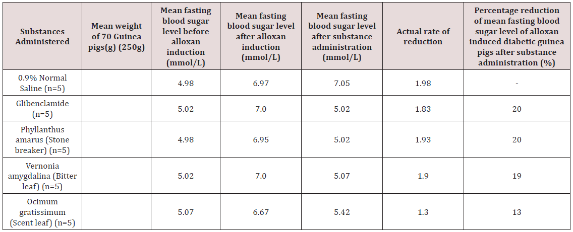
In the preliminary test, single dose alloxan induction of the guinea pigs given intraperitoneally at different concentrations of 100mg/kg, 150mg/kg and 200mg/kg body weight was seen in the Table 1 above. This Table 1 shows the mean fasting blood sugar level after a single dose alloxan induction of the five guinea pigs in three different subgroups. Induction with 200mg/kg body weight revealed an appreciable increase in the mean fasting blood sugar level of 1.6mmol/L which was greater than the readings obtained from the other two concentrations. Alloxan induction with 100mg/ kg and 150mg/kg revealed an increase in the mean fasting blood sugar of 0.4mmol/L and 0.8mmol/L respectively. These mean values were less than 1.0mmol/L. Table 2 shows the percentage reduction of the mean fasting blood sugar using aqueous leaf extracts of the three tropical herbs on alloxan induced diabetic guinea pigs when administered singly with the glibenclamide solution (oral hypoglycemic drug) as positive control and 0.9% Normal saline as a negative control. In Table 3, the percentage reduction of blood sugar level in different alloxan induced diabetic guinea pig groups were compared with that of the control groups. The percentage reduction of blood sugar level of these diabetic guinea pigs was seen to be highest for the groups of guinea pigs that had oral administration of aqueous leaf extract of Phyllanthus amarus and Vernonia amygdalina which were comparable with the positive control groups that had oral administration of glibenclamide solution. This indicates an appreciable hypoglycemic effect.
Table 1: The preliminary tests showing differential increase in mean fasting blood sugar level after alloxan induction at concentration of 100mg/kg and 200ml/kg.
Lupinepublishers-openaccess-biomedicalengineering-biosciences
Table 2: The percentage reduction of mean fasting blood sugar level readings obtained from induced groups of guinea pigs with alloxan (200mg/kg) after administration of aqueous leaf extracts using 0.9% normal saline and Globenclamide solution as negative and positive control groups respectively.
Lupinepublishers-openaccess-biomedicalengineering-biosciences
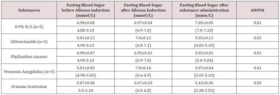
Ocimum gratissimum aqueous leaf extracts also showed moderate percentage reduction of 13% respectively which were less than the readings from positive control group of 20% reduction in mean fasting blood sugar level after those extracts. The negative control group of diabetic guinea pigs that had 0.9% Normal saline which is a physiological solution revealed no percentage reduction of blood sugar level. Using ANOVA as the statistical analytic methods, mean fasting blood sugar level of different groups as shown in this Table 3 before alloxan induction could be compared with fasting blood sugar level after alloxan induction. This Table 3 indicates that there was a significant difference between the mean fasting blood level before and after alloxan induction in all the groups (p<0.05). Also, in Table 4, Using Z-Test to compare the mean fasting blood sugar level of these guinea pigs after alloxan induction and mean fasting blood sugar after substance administration, a significant difference was seen in groups of guinea pigs that had glibenclamide solution and those that had aqueous leaf extracts of phyllanthus amarus, vernonia amygdalina and ocimum gratissimum (P<0.05). This indicates that the mean fasting blood sugar level of the groups of guinea pigs that had these aqueous leaf extracts above is comparable to the mean fasting blood sugar level of the positive control group that had glibenclamide (oral hypoglycemic agent).
Table 3: The mean fasting blood sugar level of the grouped guinea pigs before and after alloxan induction using ANOVA as the statistical analytic method as seen in the table above.
Lupinepublishers-openaccess-biomedicalengineering-biosciences
Table 4: Comparison between the mean fasting blood sugar level of the grouped guinea pigs after alloxan induction and after substance administration using Z-Test.
Lupinepublishers-openaccess-biomedicalengineering-biosciences
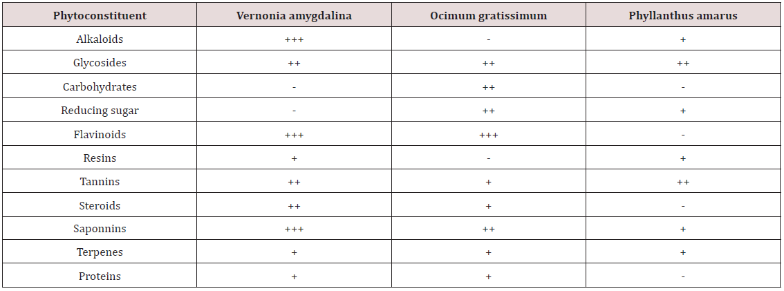
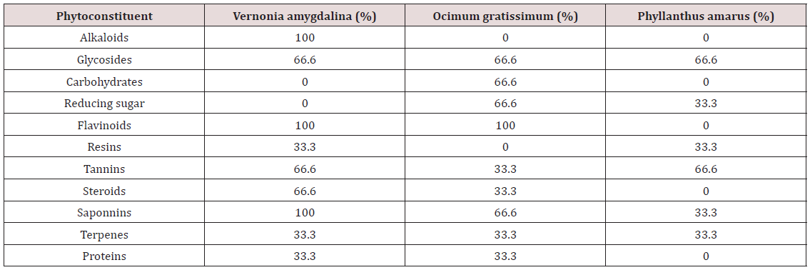
Table 5 represented above showed the quantity of the phytochemical constituents in each aqueous leaf extract of these tropical herbs during the phytochemical analysis. The presence of these phytochemical constituents is related to the hypoglycemic potency. In Table 6, the quantity of phytochemical constituents of these aqueous leaf extracts was expressed in percentages. The result obtained from the phytochemical screening of these aqueous leaf extracts as shown in Table 5 & 6 indicated that Vernonia amygdalina contains moderate levels of Alkaloids, Saponnine, Flavinoids at 100% composition and glycosides, tannins, Steroids at 66.6% composition with mild levels of Resins, terpenes and Proteins at 33.3% composition. In other words, there are more alkaloids, flavonoids, saponnins than other phytochemical compounds in vernonia amygdalina. This extract contains no carbohydrates or reducing sugars. The presence of many of these phytochemical constituents in appreciable quantities up to 100% and the absence of carbohydrate including reducing sugars may be responsible for its high hypoglycemic potency [12-25].
Table 5: The quantitative analysis obtained from phytochemical screening of the aqueous leaf extracts.
Lupinepublishers-openaccess-biomedicalengineering-biosciences
= absence of the phytochemical compound
= = presence of the phytochemical compound.
Table 6: The percentage composition of the phytochemical constituents found in phytochemical screening of the aqueous leaf extracts of these three tropical herbs used for the study.
Lupinepublishers-openaccess-biomedicalengineering-biosciences
% = percentage composition of Phytochemical constituents in extracts.
Phyllanthus amarus was found to have moderate levels of glycosides and tannins at 66.6% each, with mild level of reducing sugars, resins, saponins and terpenes at 33.3% composition. It was also found to contain no alkaloids, carbohydrates aside reducing sugars, flavonoids, steroids and proteins. The aqueous leaf extracts of this herb contain more of glycosides and tannins than any other phytochemical compounds. The saponins in this extract is far less than that of vernonia amygdalina but contains the same quantity of tannins and resins. The absence for its hypoglycemic effect. It has been noted that steroids are diabatagenic. Ocimum gratissimum contained moderate levels of flavonoids, glycosides and carbohydrates, reducing sugars and saponins at 66.6% each during phytochemical screening. The glycosides and terpenes were found to be of the same quantity as in the above two extracts. The saponins in this leaf extracts are less than the quantity obtained from vernonia amygdalina but contained the same quantity of flavonoids at 100% composition. However, flavonoids as a phytochemical constituent has antioxidation effect while saponins and peptides have antitumoral effects as stated in the early part of this work.

Conclusion

The results obtained from this study have revealed that aqueous leaf extract of phylianthus amarus, vernonia amygdalina and ocimum gratissimum have appreciable hypoglycemic effect owing to the presence of mild level of terpenes and can be termed oral hypoglycemic agents. These agents are associated with significant reduction in blood sugar level which was comparable with the effect seen in glibenclamide (positive control) and hence showed no significant difference with the effects seen in glibenclamide (p>0.05). However, using a statistical test such as Z-Test, a significant difference was seen between the post-alloxan induction and post-extract administration of these above three tropical leaves (P<0.05).

Follow on Linkedin : https://www.linkedin.com/company/lupinepublishers
Follow on Twitter   :  https://twitter.com/lupine_online


Monday, 14 January 2019

Fungus in the Feet: (LOJMS) - Lupine Publishers

Fungus in the Feet byAndrew Hague in LOJ Medical Sciences (LOJMS) - Lupine Publishers


Rich or poor, clean or dirty, infections are always attacking the feet. Here are verrucas which were treated by the local doctor with a freezing spray and ointment and for two years nothing happened until they were zapped with 100 shocks on each verruca, energy level 3 with a 20 mm focal head The verrucas have gone and new skin is taking their place. Note that only one simple treatment was needed. The verrucas are typical of what attacks those who go to swimming pools and gyms. One treatment is all it takes to kill whatever is living in the skin. Tell the patient that all their shoes must be washed or thrown away to avoid re-infection. It makes no sense to clean the feet and then get infected again as soon as they put their shoes back on The same happens with toe nail fungus (Figure 3). The fungus is killed and the nails re-grow. No drugs. No ointments. Quick and easy. It is the ability of CellSonic to kill all and any infection that makes it such an easy to use and safe tool - Lupine Publishers.

For more information click the below link
https://lupinepublishers.com/medical-science-journal/fulltext/fungus-in-the-feet.ID.000113.php
https://lupinepublishers.com/medical-science-journal/abstracts/fungus-in-the-feet.ID.000113.php
https://lupinepublishers.com/medical-science-journal/pdf/LOJMS.MS.ID.000113.pdf

For more Medical Sciences Journals click the below link
https://lupinepublishers.com/medical-science-journal/index.php

For more
Lupine Publishers Journals click the below link
https://lupinepublishers.com/


Friday, 30 November 2018

The World Politics of Dominique Moisi: (LOJMS) - Lupine publishers


The World Politics of Dominique Moisi by Thomas Scheff in LOJ Medical Sciences (LOJMS) - Lupine PublishersIn his recent (2009) book, The Geopolitics of Emotion, D. Moisi, a French political scientist, took a still somewhat unusual approach; emotions can be causal in political relationships. The book argues indeed that they may be causes of both peace and war between and within nations. He focuses on three emotions: hope, fear, and humiliation. Surprisingly, the book has received a torrent of praise from reviewers. The overwhelmingly positive response to the book is surprising because modern societies play down emotions as unimportant, even between individuals, much less nations. Most persons, even many researchers, take a materialistic stance: emotions are unimportant because both individuals and nations guide their actions toward owning money, land, resources, and other clearly visible materials. It might be that in order to write persuasively about a specific emotion, the readers must first be dissuaded from their fixed idea: emotions are unimportant [1-4] - Lupine Publishers.

For more information click the below links

Thursday, 15 November 2018

How to Evaluate Pancreaticoduodenectomy Specimens after Pancreatic Surgery? A Review: (LOJMS) Lupine publishers


Pancreaticoduodenectomy (Whipple procedure) is a complicated surgical procedure that is preferred in conditions such as malignancies and traumas of the pancreatic head, duodenum, bulb and choledoch or in case of pancreatitis. Careful attention should be paid to accurately determine the pathological and clinical stage of the disease in patients undergoing oncologic therapies that have changed over the years. This procedure, which is surgically troublesome, requires pathological sampling and care. During sampling, a pathologist must macroscopically analyse the pancreaticoduodenectomy specimens and carefully perform sampling in Lupine publishers.for more information track the below links
https://www.lupinepublishers.com/medical-science-journal/fulltext/how-to-evaluate-pancreaticoduodenectomy-specimens-after-pancreatic-surgery-a-review.ID.000104.php
https://www.lupinepublishers.com/medical-science-journal/abstracts/how-to-evaluate-pancreaticoduodenectomy-specimens-after-pancreatic-surgery-a-review.ID.000104.php
https://www.lupinepublishers.com/medical-science-journal/pdf/LOJMS.MS.ID.000104.pdf

for more informative medical sciences article click the below link
http://www.lupinepublishers.com/medical-science-journal/

For more Lupine open access journals click the below link
https://www.lupinepublishers.com/

The Body Electric: Humans Have A ‘Force Field’ Around Their Bodies

  Abstract Bioelectronic medicine (BEM) is the most recent medical revolution — not an innovation or an improvement or a step up but a radic...